Percutaneous osteoid osteoma ablation
Percutaneous destruction of an osteoid osteoma (benign painful bone tumor) by RF or cryoablation.
The information below is provided for general educational purposes only. It describes the procedure in general terms and may not apply to your specific situation. Only your interventional radiologist can provide you with personalized information adapted to your case.
What does this intervention involve?
Background
Osteoid osteoma is a benign bone tumor mainly affecting young adults, causing typical nocturnal pain relieved by aspirin. Percutaneous ablation is the gold standard.
Procedure
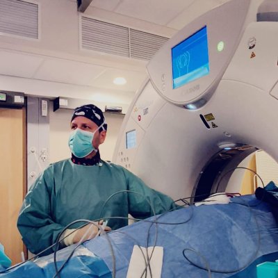
Under general anesthesia and CT guidance, a needle precisely targets the nidus. RF (90°C for 6 min) or cryoablation destroys the tumor. Duration: 30-60 minutes.
Risks
Fracture (rare), skin burn, nerve injury (spinal location), recurrence (5%).
Recovery
Same-day discharge. Pain resolution within days. No routine follow-up imaging.
Practical information
General anesthesia. Hospital stay: one to two nights.
This information does not replace a medical consultation. Each procedure is adapted to the patient's individual situation. Your doctor will explain the specific details, expected benefits and potential risks during your consultation.
Doctors and centers/departments performing this intervention
12 doctors
CHU CHARLEROI
CHU (Centre Hospitalier Universitaire)Charleroi
CHU Helora Site Jolimont
CHU (Centre Hospitalier Universitaire)La Louvière

Dr Alexia DABADIE
Radiologue interventionnelDr Arthur DAVID
Radiologue interventionnelNantes
Dr Vincent DUROUS
Radiologue interventionnelAnnecy, Argonay, Épagny
Dr Cédric FOUSSIER
Radiologue interventionnelParis
Hôpital Européen Marseille
Clinique privéeMarseille
Dr Lucas MOSCATELLI
Radiologue interventionnelSaint-Paul
Dr Matthieu PAPILLARD
Radiologue interventionnelGenève
RIVA - Hôpital Privé Océane Vannes
Clinique privéeVannes
Service de radiologie interventionnelle - CHU de NANTES
CHU (Centre Hospitalier Universitaire)Nantes
Dr Nicolas VILLARD
Radiologue interventionnelLausanne