Lesion localization before surgery (guide wire, hook, clip)
Percutaneous placement of a marker (wire, clip, dye) in or near a tumor to guide the surgeon during the operation.
The information below is provided for general educational purposes only. It describes the procedure in general terms and may not apply to your specific situation. Only your interventional radiologist can provide you with personalized information adapted to your case.
What does this intervention involve?
Background and indications
When a tumor is small, deep or not palpable, localization is needed before surgery so the surgeon can precisely identify it. This frequently concerns lung nodules, liver lesions or other deep tumors.
Benefits
Enables more precise and conservative surgery, avoiding removal of excessive healthy tissue.
Procedure
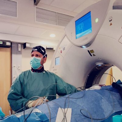
Under local anesthesia, a guide wire (hook), clip or dye is placed in or near the lesion under CT or ultrasound guidance. The procedure is quick (15-30 minutes) and performed the same day or day before surgery.
Risks
Pneumothorax (for lung lesions), minor puncture site bleeding, marker migration (rare).
Recovery and follow-up
Surgery is performed within hours. The marker is removed with the surgical specimen.
Practical information
Local anesthesia. Outpatient procedure (return home the same day).
This information does not replace a medical consultation. Each procedure is adapted to the patient's individual situation. Your doctor will explain the specific details, expected benefits and potential risks during your consultation.
Doctors and centers/departments performing this intervention
11 doctors

Centre Hospitalier de Saint-Quentin
Centre hospitalierSaint-Quentin
CHU Helora Site Jolimont
CHU (Centre Hospitalier Universitaire)La Louvière

Dr Frederic COHEN
Radiologue interventionnelAubagne, Marseille
Dr Arthur DAVID
Radiologue interventionnelNantes
Hôpital Européen Marseille
Clinique privéeMarseille

Dr Thibaud LEFORT
Radiologue interventionnelLyon

Dr Matthieu PAPILLARD
Radiologue interventionnelGenève
RIVA - Hôpital Privé Océane Vannes
Clinique privéeVannes

Service de radiologie interventionnelle - CHU de NANTES
CHU (Centre Hospitalier Universitaire)Nantes

Service de radiologie interventionnelle - Hôpital de Valenciennes
Centre hospitalierValenciennes

Dr Nicolas VILLARD
Radiologue interventionnelLausanne