Lipiodol CT scan
Diagnostic examination combining Lipiodol injection into hepatic arteries with CT scanning to detect and characterize liver tumors.
The information below is provided for general educational purposes only. It describes the procedure in general terms and may not apply to your specific situation. Only your interventional radiologist can provide you with personalized information adapted to your case.
What does this intervention involve?
Background and indications
Lipiodol-CT is used to detect small liver tumors that are difficult to visualize on standard imaging, particularly before chemoembolization or to complete hepatocellular carcinoma staging.
Benefits
Highly sensitive detection of small hepatic tumors by exploiting preferential Lipiodol uptake in tumor tissue.
Procedure
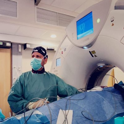
A catheter is introduced into the femoral or radial artery and guided to the hepatic artery. Lipiodol is injected, then a CT scan is performed in the following hours or days to observe its distribution in the liver. The examination lasts about 1 hour.
Risks
Risks are those of arteriography (puncture site hematoma, contrast allergy) and are generally low.
Recovery and follow-up
Same-day or next-day discharge. Results guide the therapeutic strategy.
Practical information
Local anesthesia. Outpatient procedure (return home the same day).
This information does not replace a medical consultation. Each procedure is adapted to the patient's individual situation. Your doctor will explain the specific details, expected benefits and potential risks during your consultation.
Doctors and centers/departments performing this intervention
4 doctors
CHU Helora Site Jolimont
CHU (Centre Hospitalier Universitaire)La Louvière

Dr Frederic COHEN
Radiologue interventionnelAubagne, Marseille

Dr Vincent DUROUS
Radiologue interventionnelAnnecy, Argonay, Épagny

Dr Nicolas VILLARD
Radiologue interventionnelLausanne